Comprehensive eye examination is performed by a specialist ophthalmologist, with the assistance of a nurse or medical technician. A standard ophthalmologic examination lasts up to 60 minutes and is completely comfortable, safe and painless for the patient.
What does a comprehensive eye examination include?
The basic (general) ophthalmologic examination starts with a conversation with the patient (taking the medical history) about the symptoms the patient is experiencing, previous illnesses and other important questions. After the history is taken, the examination is performed and includes:
Determining visual acuity for near and distance
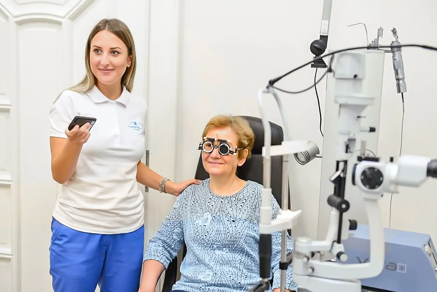
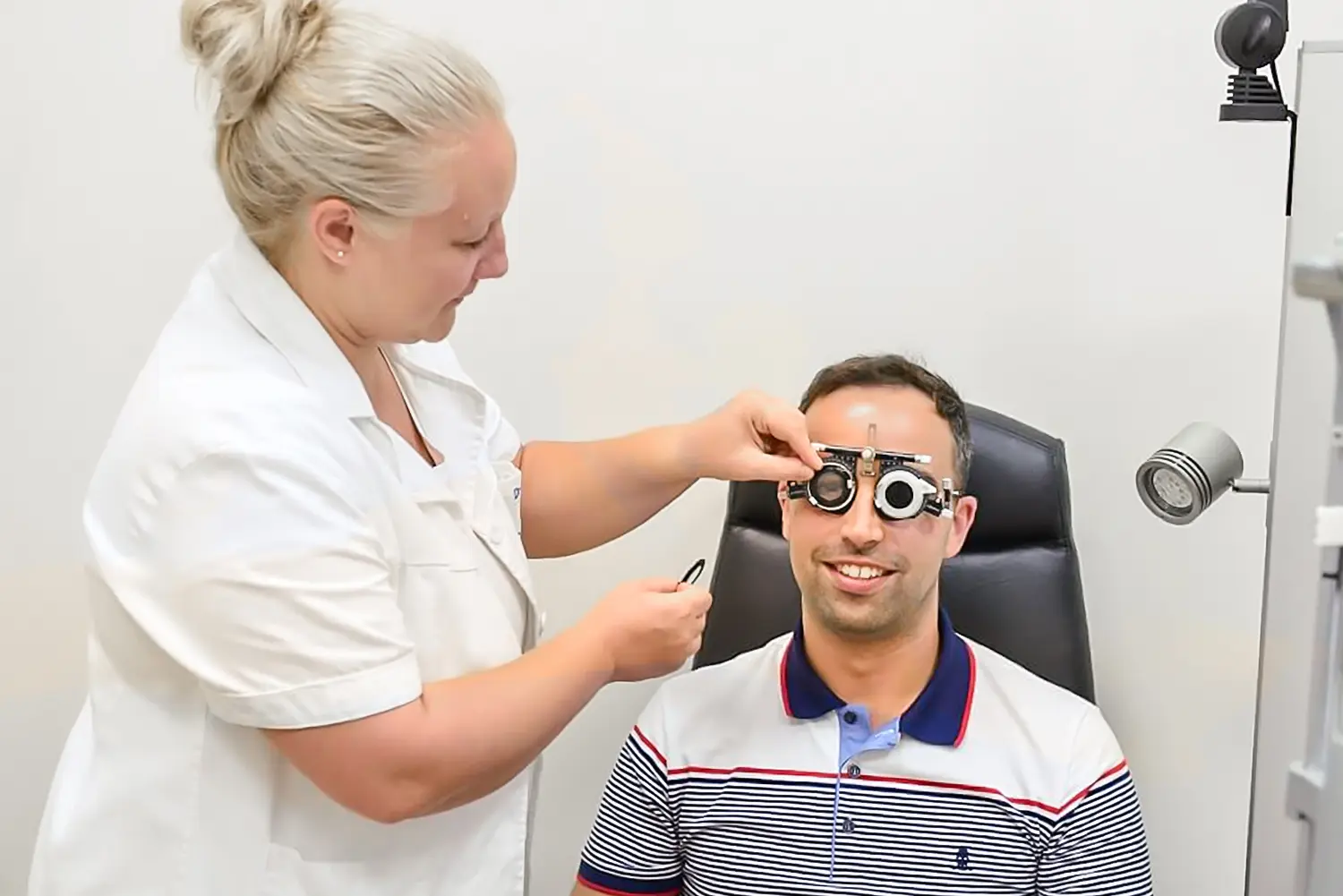
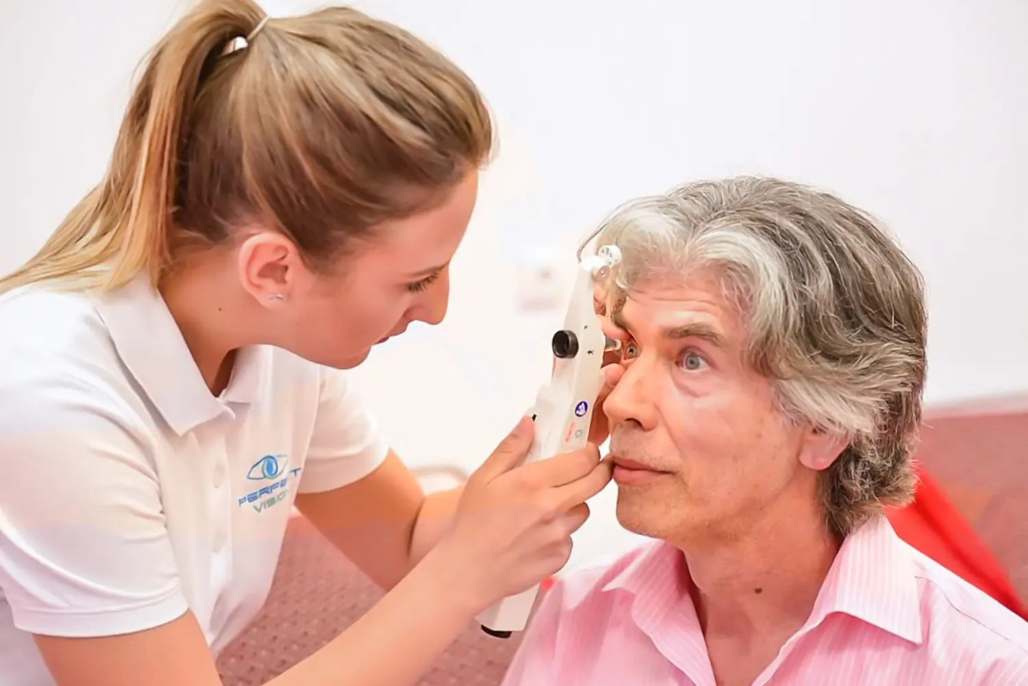
The eye examination begins by checking the refractive error (diopters) with undilated pupils using an automatic keratorefractometer. If needed, corneal topography is also performed, which provides detailed corneal maps, its refractive power and any irregularities.
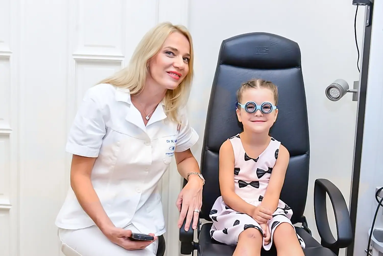
During subjective refraction, the patient reads letters or numbers (children look at pictures) from a visual acuity chart, while the doctor uses spherical or cylindrical lenses to correct myopia, hyperopia or astigmatism.
In children and young patients, because of accommodation, the examination is often performed in cycloplegia (with dilated pupils), where drops temporarily block the lens accommodation so that the exact diopter value can be determined.
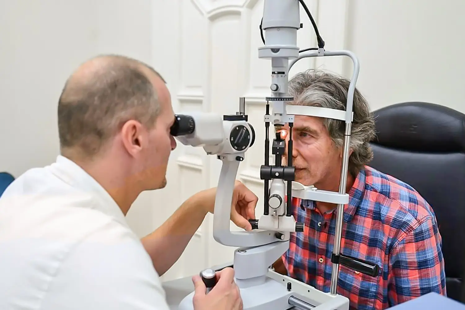
Examination of the anterior segment of the eye
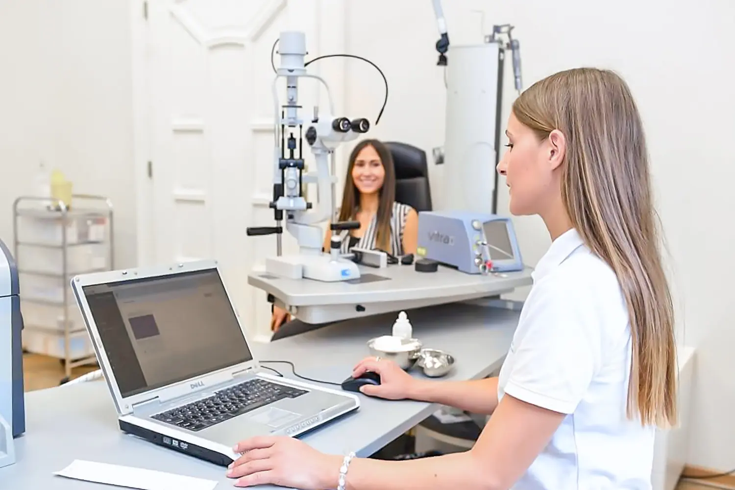
The anterior segment of the eye includes the cornea, conjunctiva, sclera, iris, anterior chamber, lens and the anterior part of the vitreous body. Using a device called a slit lamp (biomicroscope), the ophthalmologist illuminates and examines each part of the anterior segment of the eye under high magnification.
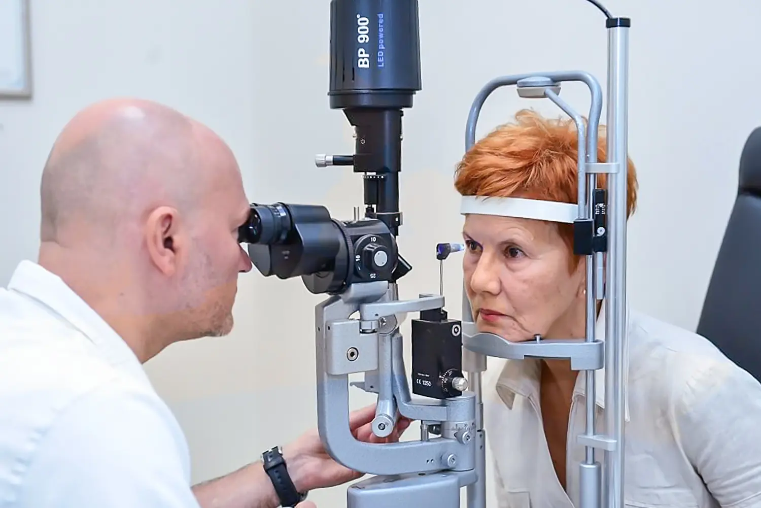
Fundus examination
The ocular fundus is the inner part of the eye located behind the lens. The most complete examination is performed with dilated pupils and includes the retina with its structures: the optic disc (optic nerve head), macula and retinal blood vessels.
It is especially important in patients with diabetes, patients with high blood pressure and in highly myopic patients, where regular examinations are essential for timely diagnosis and treatment.
Examination of the anterior segment of the eyeFundus examination
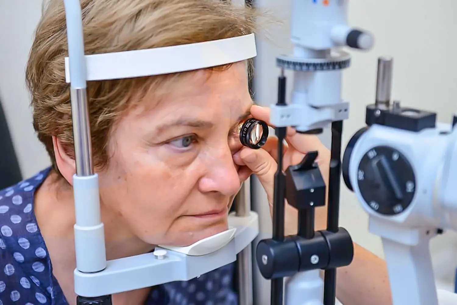
Measuring intraocular pressure
Before measuring intraocular pressure, the surface of the eye is anesthetized with eye drops, after which an applanation tonometer is gently applied to the cornea to show the pressure values. Normal values range between 10 and 20 mmHg.
If the eye is healthy and there is only a refractive error, eyeglasses or contact lenses are prescribed during this examination.
Timely and comprehensive eye examination allows detection of eye diseases at an early stage, even when there are no visible symptoms (cataract, glaucoma, diabetic retinopathy, macular degeneration, etc.). If needed, recommendations for further treatment and referral to a subspecialist are provided.
Who performs an eye examination?
Differences between an ophthalmologist, optometrist and optician and when to see each of them.
Pronađite odgovore na najčešća pitanja o laserskom skidanju dioptrije, katarakti i ugradnji sočiva. Sve što treba da znate pre nego što donesete odluku o zahvatu.
Ne, zahvat je potpuno bezbolan. Traje svega nekoliko minuta, a već sutradan možeš normalno da funkcionišeš bez naočara ili sočiva.
Ako primećuješ da ti je vid zamućen, boje blede i noću teško voziš, to su jasni znakovi. Operacija vraća bistrinu vida i ne odlaže se kad se jednom dijagnostikuje.
Najveća prednost je što zaboravljaš na naočare – i za blizinu i za daljinu. To znači da čitaš knjigu, koristiš telefon i voziš bez dodatnih pomagala.
Itekako imaš! Za to postoje torična sočiva, koja su posebno napravljena da isprave astigmatizam i daju jasan vid.
To su veštačka sočiva koja se ubacuju u oko, ali tvoje prirodno sočivo ostaje netaknuto. Odlična su za mlađe ljude sa visokom dioptrijom koji nisu kandidati za laser.